
توضیح محصول
———————————————————
دانلود کتاب الکترونیکی
———————————————————
 How to Buy
Sectional Anatomy by MRI and CT TRUE PDF price 1€
you should pay 1€ Euro via below payment Link
https://yekpay.io/en/baharmedbook.com
Pay attention to Send Money To abbas kouhiesfahani baharmedbook_manager_Bank account
After Payment confirmation ,contact us by shopping guide on WhatsApp or info@baharmedbook.com
and You will Receive Direct Download Link
———————————————————
Product Description
———————————————————
Download e-book
———————————————————
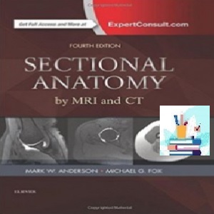
Sectional Anatomy by MRI and CT
TRUE PDF
Price 1€
2017
(Original quality)
4th Edition
Now available with state-of-the-art digital enhancements, the highly anticipated 4th edition of this classic reference is even more relevant and accessible for daily practice. A sure grasp of cross sectional anatomy is essential for accurate radiologic interpretation, and this atlas provides exactly the information needed in a practical, quick reference format. New color coding of anatomic structures and new scroll and zoom capabilities on photos in the eBook version make this title an essential diagnostic tool for both residents and practicing radiologists.
- Expert Consult eBook version included with purchase. This enhanced eBook experience allows you to search all of the text, figures, images, and references from the book on a variety of devices.
- Color-coded labels for nerves, vessels, muscles, bone tendons, and ligaments facilitate accurate identification of key anatomic structures.
- Scroll and zoom capabilities on photos in the accompanying eBook version enable easier accessibility during interpretation sessions and real-time resident education.
- Carefully labeled MRIs for all body parts, as well as schematic diagrams and concise statements, clarify correlations between bones and tissues.
- CT scans for selected body parts enhance anatomic visualization.
- More than 2,300 state-of-the-art images can be viewed in three standard planes: axial, coronal, and sagittal.
About the Author
Mark W. Anderson, MD, Harrison Distinguished Teaching Professor of Radiology; Chief, Musculoskeletal Imaging, Professor of Orthopaedic Surgery, University of Virginia, Charlottesville, Virginia
Michael G Fox, MD, Associate Professor of Radiology and Medical Imaging, Associate Professor of Orthopaedic Surgery, University of Virginia, Charlottesville, Virginia
Contents
۱ MRI of the Pectoral Girdle and Chest Wall
۲ MRI of the Shoulder
۳ MR Arthrography of the Shoulder
۴ MRI of the Anri
۵ MRI of the Elbow
۶ MRI of the Forearm
۷ MRI of the Wrist
۸ MRI of the Hand
۹ MRI of the Hip
۱۰ MR Arthrography of the Hip
۱۱ MRI of the Thigh
۱۲ MRI of the Knee
۱۳ MRI of the Leg
۱۴ MRI of the Ankle
۱۵ MRI of the Foot
۱۶ MRI of the Thoracic Spine
۱۷ MRI of the Lumbar Spine
۱۸ CT of the Thorax
۱۹ MRI of the Heart
۲۰ CT of the Abdomen
۲۱ MRI of the Abdomen
۲۲ CT of the Male Pelvis
۲۳ CT of the Female Pelvis
۲۴ MRI of the Male Pelvis
۲۵ MRI of the Female Pelvis