
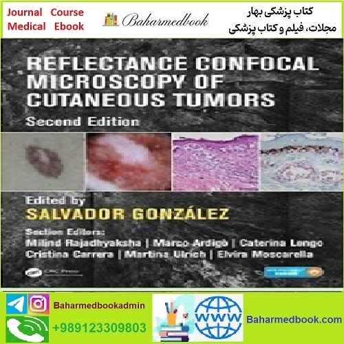
توضیح محصول
———————————————————
دانلود کتاب الکترونیکی
———————————————————
میکروسکوپ کانفوکال بازتاب تومورهای پوستی
پی دی اف واقعی
قیمت 1 یورو
2017
(کیفیت اصلی)
ویرایش 2
میکروسکوپ کانفوکال انعکاس ضایعات پوست را قادر می سازد بدون برش، اما با دقت تشخیصی بهبود یافته، ارزیابی همبستگی درموسکوپی-هیستولوژیک، ارزیابی حاشیه های جراحی و همچنین سرعت و راحتی برای پزشک و بیمار بررسی شوند. این متن بهطور گسترده بزرگشده و بهروز شده، وضعیت فعلی و آینده هنر را برای کسانی که با تشخیص و درمان تومورهای پوستی مرتبط هستند، با مقدار زیادی افزایش یافته در مورد الگوهای طبیعی پوست در طول زندگی و تومورهای غیرملانوسیتی بررسی میکند.
امکانات
دارای رنگ در سراسر
دسترسی به کتاب الکترونیکی یکپارچه برای سهولت مرجع دارد
مواد روی الگوهای طبیعی پوست مورد انتظار در طول زندگی را به شدت افزایش داده است
مواد روی تومور غیرملانسیتیک را بسیار افزایش داده است
درباره نویسنده
سالوادور گونزالس هم در دانشگاه آلکالا و هم در بیمارستان رامون وای کخال، مادرید، اسپانیا و مرکز سرطان مموریال اسلون کترینگ، نیویورک، ایالات متحده، قرار ملاقات دارد. رئیس کنونی انجمن بین المللی میکروسکوپ کانفوکال، او در تحقیقات و عمل بالینی در سطح بین المللی فعال است.
فهرست
۱. مبانی میکروسکوپ کانفوکال بازتاب
۲. کاربرد کانفوکال در تمرین روزمره
۳. روش های محاسباتی در میکروسکوپ کانفوکال پوست
۴. نقش تله درماتولوژی در میکروسکوپ کانفوکال بازتابی
۵. دستگاه میکروسکوپ کانفوکال مینیاتوری برای تصویربرداری از پوست
۶. میکروبیوپسی های هدایت شده با میکروسکوپ کانفوکال انعکاسی برای تجزیه و تحلیل مولکولی هدفمند
۷. نوار موزاییک
۸. میکروسکوپ کانفوکال چندوجهی برای سرطانهای پوست غیرملانومی در شرایط خارج از بدن
۹. میکروسکوپ کانفوکال انعکاس حین عمل برای هدایت بالقوه جراحی Mohs و سایر جراحی های پوستی
۱۰. حذف لیزری کارسینوم سلول بازال با هدایت میکروسکوپ کانفوکال
۱۱. پوست معمولی: اصطلاحات
۱۲. ساختارهای آدنکس و حسی پوست
۱۳. تغییرات توپوگرافی و فوتوتیپ پوست با تاکید ویژه بر پوست صورت و آکرال
۱۴. بلوغ پوست نوزاد: تغییرات ساختاری آشکار شده توسط میکروسکوپ کانفوکال بازتابی in vivo و چشم اندازهای آینده
۱۵. پیری پوستی: توضیحات و الگوریتم ها
۱۶. مخاط دهان و تناسلی سالم
۱۷. تشخیص RCM نئوپلاسم های ملانوسیتی: اصطلاحات، الگوریتم ها و دقت و ادغام بالینی آنها
۱۸. ارتباط بافت شناسی RCM در ضایعات ملانوسیتی
۱۹. خال های معمولی
۲۰. خال های ملانوسیتی مادرزادی
۲۱. خال های اسپیتز رید
۲۲. خال های دیسپلاستیک
۲۳. چگونه صفات ژنتیکی ممکن است بر مورفولوژی درموسکوپی و کانفوکال خال ها تأثیر بگذارد
۲۴. گسترش سطحی و ملانوم ندولار (شامل ملانوم آملانوتیک)
۲۵. Lentigo maligna و lentigo maligna melanoma
۲۶. خال ها و ملانوما
۲۷. ملانوما در مکان های خاص
۲۸. ضایعات که ساختارهای پسرونده را آشکار می کنند
۲۹. اصطلاحات، تجزیه و تحلیل الگو و الگوریتم های RCM به کار رفته در تومورهای غیر ملانوسیتی
۳۰. میکروسکوپ کانفوکال بازتاب – همبستگی های بافت شناسی برای تومورهای غیر ملانوسیتی
۳۱. الگوهای عروقی در تومورهای غیر ملانوسیتی
۳۲. کراتوز اکتینیک پیگمانته و پوروکراتوزیس
۳۳. نئوپلازی سنگفرشی (زیر انواع) و پیشرفت
۳۴. کارسینوم سلول بازال: انواع فرعی
۳۵. شبیه سازهای کارسینوم سلول بازال: تریکواپیتلیومای پورومار و پاپول های فیبری صورت
۳۶. هیپرپلازی سباسه و آدنوم. آکانتوما و درماتوفیبروم سلولی شفاف
۳۷. لنتیگو خورشیدی و کراتوز شبیه لیکن پلان
۳۸. کراتوز سبورئیک: سرنخ های اصلی برای جلوگیری از تشخیص اشتباه شبیه سازهای شبیه کراتوز سبورئیک
۳۹. میکروسکوپ کانفوکال انعکاس in vivo برای تشخیص و مدیریت بیماری پاژه خارج پستانی
40. تومورها و ناهنجاری های عروقی خوش خیم و سارکوم کاپوزی
۴۱. Mycosis fungoides و سایر لنفوم های پوستی سلول T
۴۲. دقت تشخیصی RCM در یک محیط بالینی
۴۳. ادغام میکروسکوپ کانفوکال بازتاب برای مدیریت بیماران مبتلا به خال های متعدد
۴۴. لنتیگو بدخیمی ملانوم: نظارت بر درمان غیر تهاجمی و نقشه برداری حاشیه قبل از جراحی
۴۵. مدیریت تومور ناخن با میکروسکوپ کانفوکال حین عمل
۴۶. سرطان میدانی و نظارت بر درمان
۴۷. پایش درمان غیرتهاجمی کارسینوم سلول بازال
۴۸. میکروسکوپ کانفوکال فلورسنت Ex vivo برای هدایت جراحی میکروگرافیک Mohs

How to Buy
Reflectance Confocal Microscopy of Cutaneous Tumors TRUE PDF price 1€
you should pay 1€ Euro via below payment Link
https://yekpay.io/en/baharmedbook.com
Pay attention to Send Money To abbas kouhiesfahani baharmedbook_manager_Bank account
After Payment confirmation ,contact us by shopping guide on WhatsApp or info@baharmedbook.com
and You will Receive Direct Download Link
———————————————————
Product Description
———————————————————
Download e-book
———————————————————
Reflectance Confocal Microscopy of Cutaneous Tumors
TRUE PDF
Price 1€
2017
(Original quality)
2th Edition
Reflectance confocal microscopy enables lesions in skin to be examined without excision, but with improved diagnostic accuracy, assessment of dermoscopic-histologic correlation, assessment of surgical margins, as well as speed and convenience for the physician and patient. This extensively enlarged and updated text reviews the current and future state of the art for those involved with the diagnosis and treatment of skin tumors, with a greatly increased amount of material on the expected normal patterns of skin throughout life and on non-melanocytic tumors.
Features
features color throughout
has integral ebook access for ease of reference
has greatly increased material on the expected normal patterns of skin throughout life
has greatly increased material on non melancytic tumor
About the Author
Salvador Gonzalez has dual appointments at both Alcalá University and Hospital Ramon y Cajal, Madrid, Spain, and Memorial Sloan-Kettering Cancer Center, New York, USA. The current President of the International Society of Confocal Microscopy, he is active in research and clinical practice internationally.
Contents
۱. Fundamentals of reflectance confocal microscopy
۲. Confocal application in everyday practice
۳. Computational methods in skin confocal microscopy
۴. The role of teledermatology in reflectance confocal microscopy
۵. Miniature confocal microscopy devices for imaging skin
۶. Reflectance confocal microscopy-guided microbiopsies for targeted molecular analysis
۷. Strip-mosaicking
۸. Multimodal confocal microscopy for nonmelanoma skin cancers ex vivo
۹. Intraoperative reflectance confocal microscopy to potentially guide Mohs surgery and other dermatologic surgeries
۱۰. Laser ablation of basal cell carcinoma guided by confocal microscopy
۱۱. Normal skin: Terminology
۱۲. Adnexal and sensory structures of the skin
۱۳. Topographic and skin phototype variations of skin with special emphasis on facial and acral skin
۱۴. Infant skin maturation: Structural changes revealed by in vivo reflectance confocal microscopy and future perspectives
۱۵. Cutaneous photoaging: Description and algorithms
۱۶. Healthy oral and genital mucosa
۱۷. RCM diagnosis of melanocytic neoplasms: Terminology, algorithms and their accuracyr and clinical integration
۱۸. RCM-histology correlation in melanocytic lesions
۱۹. Common nevi
۲۰. Congenital melanocytic nevi
۲۱. Spitz-Reed nevi
۲۲. Dysplastic nevi
۲۳. How genetic traits may influence the dermoscopic and confocal morphology of nevi
۲۴. Superficial spreading and nodular melanoma (including amelanotic melanoma)
۲۵. Lentigo maligna and lentigo maligna melanoma
۲۶. Nevi and melanoma
۲۷. Melanoma in special locations
۲۸. Lesions revealing regressive structures
۲۹. Terminology, pattern analysis and algorithms of RCM applied to nonmelanocytic tumors
۳۰. Reflectance confocal microscopy— Histology correlations for nonmelanocytic tumors
۳۱. Vascular patterns in nonmelanocytic tumors
۳۲. Pigmented actinic keratosis and porokeratosis
۳۳. Squamous neoplasia (subtypes) and progression
۳۴. Basal cell carcinoma: Subtypes
۳۵. Basal cell carcinoma simulators: Poromar trichoepithelioma and fibrous papules of the face
۳۶. Sebaceous hyperplasia and adenoma. Clear cell acanthoma and dermatofibroma
۳۷. Solar lentigo and lichen planus-like keratosis
۳۸. Seborrheic keratosis: The main clues to avoid misdiagnosing seborrheic keratosis-like simulators
۳۹. In vivo reflectance confocal microscopy for diagnosis and management of extramammary Paget disease
۴۰. Benign vascular tumors and malformations,, and Kaposi sarcoma
۴۱. Mycosis fungoides and other cutaneous T-cell lymphomas
۴۲. Diagnostic accuracy of RCM in a clinical setting
۴۳. Integration of reflectance confocal microscopy for the management of patients with multiple nevi
۴۴. Lentigo maligna melanoma: Monitoring of noninvasive treatment and margin mapping prior to surgery
۴۵. Nail tumor management by intraoperative confocal microscopy
۴۶. Field cancerization and monitoring of treatment
۴۷. Monitoring of noninvasive treatment of basal cell carcinoma
۴۸. Ex vivo fluorescent confocal microscopy to guide micrographic Mohs surgery