
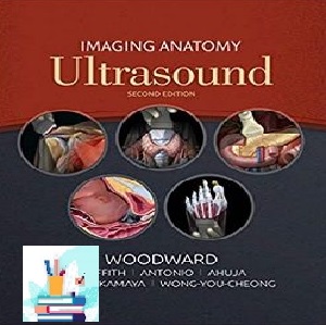
توضیح محصول
———————————————————
دانلود کتاب الکترونیکی
———————————————————
سونوگرافی آناتومی تصویربرداری
پی دی اف واقعی
قیمت 1 یورو
2018
(کیفیت اصلی)
ویرایش 2
طراحی شده برای کمک به یادگیری سریع یا مرور آناتومی طبیعی و تأیید انواع آن، تصویربرداری آناتومی: اولتراسوند، ویرایش دوم، مرجع نهایی در سراسر جهان است که شما را در زمینه تغییر سریع تصویربرداری اولتراسوند از طریق پوشش جامع آناتومی سونوگرافی برای سر و گردن، آشنا میکند. ، اسکلتی عضلانی، شکم و لگن، مامایی و جنین شناسی، سر نوزاد و عروقی. با بهروزرسانی بیشتر تصاویر، این نسخه دوم کاملاً بهروز و با تصویرسازی بالا است، که وقتی با ساختاری منظم و قابل پیگیری آسان ترکیب میشود، این عنوان منحصربهفرد را در زمینه خود بینظیر میکند.
در هر ناحیه تشریحی که در آن از سونوگرافی استفاده میشود، مرجع تخصصی در محل مراقبت ارائه میشود
تصاویر دارای برچسب غنی را همراه با تفسیر مرتبط و همچنین تصاویر پیشاهنگی ریز برای نشان دادن قرارگیری مبدل ارائه میکند.
دارای مجموعهای قوی از همبستگیهای CT/MR است که اهمیت تصویربرداری چندوجهی را در عملکرد بالینی مدرن برجسته میکند.
منعکس کننده پیشرفت های چشمگیر اخیر در تجهیزات و تکنیک ها با تصاویر پیشرفته در سراسر
شامل بخش اسکلتی عضلانی گسترده، محتوای OB/GYN جدید و توسعه یافته شامل کف لگن، و پوشش جدید سونوگرافی سه بعدی
درباره نویسنده
پروفسور جیمز اف گریفیث یک رادیولوژیست بالینی دانشگاهی است که در دانشگاه چینی هنگ کنگ کار می کند. او 25 سال تجربه کار با سونوگرافی اسکلتی عضلانی را در یک محیط بالینی از آغاز اولیه سونوگرافی با وضوح بالا تا عمل بالینی دارد. او رئیس یک واحد تصویربرداری اسکلتی عضلانی شلوغ است که تمام جنبه های تصویربرداری اسکلتی عضلانی از جمله سونوگرافی، CT، MRI، PET-CT و مداخله را پوشش می دهد. این یکپارچگی متقاطع تصویربرداری به او اجازه میدهد تا به طور کامل از مزایای اولتراسوند در کار و درمان بیماریهای مختلف اسکلتی عضلانی قدردانی کند. او نزدیک به 300 مقاله با داوری در مورد جنبه های مختلف تصویربرداری اسکلتی عضلانی و همچنین انجام و شرکت در حداقل چهل کارگاه اولتراسوند اسکلتی عضلانی منتشر کرده است.
فهرست
بخش 1: مغز و ستون فقرات
بخش 2: سر و گردن
بخش 3: قفسه سینه
بخش 4: شکم
بخش 5: لگن
بخش 6: اندام فوقانی
بخش 7: اندام تحتانی
بخش 8: زنان و زایمان و آناتومی رشد

How to Buy
Imaging Anatomy Ultrasound TRUE PDF price 1€
you should pay 1€ Euro via below payment Link
https://yekpay.io/en/baharmedbook.com
Pay attention to Send Money To abbas kouhiesfahani baharmedbook_manager_Bank account
After Payment confirmation ,contact us by shopping guide on WhatsApp or info@baharmedbook.com
and You will Receive Direct Download Link
———————————————————
Product Description
———————————————————
Download e-book
———————————————————
Imaging Anatomy Ultrasound
TRUE PDF
Price 1€
2018
(Original quality)
2th Edition
Designed to help you quickly learn or review normal anatomy and confirm variants, Imaging Anatomy: Ultrasound, second edition, is the ultimate reference worldwide, keeping you current within the fast-changing field of ultrasound imaging through comprehensive coverage of sonographic anatomy for head and neck, musculoskeletal, abdomen and pelvis, obstetrics and embryology, neonatal head, and vascular. With most images updated, this second edition is completely up-to-date and highly illustrated, which when combined with an orderly, easy-to-follow structure, make this unique title unmatched in its field.
-
- Provides expert reference at the point of care in every anatomical area where ultrasound is used
- Presents richly labeled images with associated commentary as well as thumbnail scout images to show transducer placement
- Features a robust collection of CT/MR correlations, highlighting the importance of multimodality imaging in modern clinical practice
- Reflects the recent dramatic improvements in equipment and techniques with state-of-the-art images throughout
- Includes an expanded musculoskeletal section, new and expanded OB/GYN content including pelvic floor, and new coverage of 3D ultrasound
About the Author
Professor James F Griffith is an academic clinical radiologist working at The Chinese University of Hong Kong. He has 25 years’ experience working with musculoskeletal ultrasound in a clinical setting from the initial inception of high-resolution ultrasound into clinical practice. He is a Head of a busy Musculoskeletal Imaging Unit covering all aspects of musculoskeletal imaging including ultrasound, CT, MRI, PET-CT and intervention. This imaging cross-integration allows him to fully appreciate the benefits of ultrasound in the work up and treatment of different musculoskeletal conditions. He has published nearly 300 peer-reviewed papers on various aspects of musculoskeletal imaging as well as conducting and participating in at least forty musculoskeletal ultrasound workshops.
Contents
SECTION 1: Brain and Spine
SECTION 2: Head and Neck
SECTION 3: Thorax
SECTION 4: Abdomen
SECTION 5: Pelvis
SECTION 6: Upper Extremity
SECTION 7: Lower Extremity
SECTION 8: Obstetrics and Developmental Anatomy