0

فروش

30000

تومان

/5
0 دیدگاه

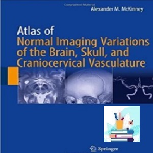
Atlas of Normal Imaging Variations of the Brain Skull and Craniocervical Vasculature TRUE PDF price 1€

حجم فایل

نرم افزار

پلاگین مورد نیاز

تاریخ به روزرسانی

خریداران محصول

پشتیبانی

توضیح محصول
معرفی Atlas of Normal Imaging Variations of the Brain Skull and Craniocervical Vasculature TRUE PDF price 1€

توضیح محصول

———————————————————

دانلود کتاب الکترونیکی

———————————————————

اطلس تغییرات تصویربرداری طبیعی از جمجمه مغز و عروق کرانیوسرویکال


پی دی اف واقعی


قیمت 1 یورو


2017


(کیفیت اصلی)


ویرایش 1

این اطلس تغییرات تصویربرداری طبیعی از مغز، جمجمه و عروق کرانیوسرویکال را ارائه می دهد. تصویربرداری رزونانس مغناطیسی (MR) و توموگرافی کامپیوتری (CT) در 10 سال گذشته پیشرفت چشمگیری داشته است، به ویژه در مورد تکنیک های جدید و تصویربرداری سه بعدی. یکی از مشکلات عمده ای که توسط رادیولوژیست ها و پزشکان تجربه می شود، تفسیر انواع نرمال در مقایسه با ناهنجاری هایی است که واریانت ها تقلید می کنند. از طریق مجموعه گسترده ای از تصاویر، این کتاب طیفی از ظاهرها را برای هر نوع با تصویربرداری سه بعدی همراه برای تأیید ارائه می دهد. مصنوعات رایج در MR و CT که بیماری را شبیه‌سازی می‌کنند را بررسی می‌کند. هر یک از انواع را از نظر آناتومی مربوطه مورد بحث قرار می دهد. و موارد مقایسه ای را به منظور تشخیص یافته های طبیعی از ناهنجاری ها ارائه می کند. این شامل انواع رایج و همچنین انواع تازه شناسایی شده است که با تکنیک های اخیراً توسعه یافته مانند تصویربرداری با وزن انتشار و CT چند آشکارساز/چند تکه تجسم شده اند. این کتاب همچنین انواع طبیعی تصویربرداری در موارد کودکان را برجسته می کند.

Atlas of Normal Imaging Variations of Brain, Skull, and Craniocervical Vasculature منبع ارزشمندی برای رادیولوژیست های عصبی، متخصصان مغز و اعصاب، جراحان مغز و اعصاب و رادیولوژیست ها در تفسیر رایج ترین و قابل شناسایی ترین انواع و استفاده از بهترین روش ها برای طبقه بندی مناسب آنها است.

درباره نویسنده

الکساندر ام. مک‌کینی، MD، بخش رادیولوژی، دانشگاه مینه‌سوتا، مینیاپولیس، MN، ایالات متحده

فهرست

۱. مغز. معرفی
۲. اکتوپی لوزه مخچه
۳. فلوکولوس کاذب مخچه
۴. کیست های آراکنوئید مگا سیسترنا مگنا و رتروسربلیار
۵. عصب جمجمه ای VII. افزایش کنتراست طبیعی در تصویربرداری تشدید مغناطیسی
۶. عدم تقارن برجسته غار مکل و سفالوسل راس پتروس
۷. چربی سینوس غار و شبه توده
۸. هیپوفیز Variationsr Artifacts، Primary Empty Sella و Incidentalmas
۹. غشای Liliequist
۱۰. غده پینه آل. اندازه معمولی، ظاهر، و افزایش
۱۱. شبکه کوروئید. مکان ها و ظواهر عادی
۱۲. هیپوکامپی، دم دمی و انواع اپاندیمال
۱۳. انواع خط میانی Septum Pellucidum، Corpus Callosum و Massa Intermedia
۱۴. فضاهای پریوسکولار گشاد شده
۱۵. بزرگ شدن یا عدم تقارن بطن های جانبی شبیه سازی هیدروسفالی
۱۶. کاهش حجم مغزی یا مخچه شبیه سازی هماتوم ساب دورال، هیگروما و کیست آراکنوئید
۱۷. کلسیفیکاسیون های دورال مکان ها و ظواهر عادی
۱۸. تقویت کنتراست شبه لپتومننژیال
۱۹. گانگلیون پایه. کلسیفیکاسیون فیزیولوژیک
۲۰. تصویربرداری با وزن حساس. مفاهیم، تنوع گانگلیون پایه در رسوب آهن مرتبط با سن، و مصنوعات
۲۱. آهسته جریان، ناهنجاری های عروقی بدون علامت. تلانژکتازی های مویرگی مغز و ناهنجاری های وریدی رشدی
۲۲. ضایعات کاذب MRI مغز. 3.0 T Imaging، FLAIR و Diffusion-Weighted Imaging
۲۳. مغز کودکان. تغییرات طبیعی در رشد، بلوغ و میلیناسیون
۲۴. جمجمه. معرفی
۲۵. جمجمه کودکان. بخیه های طبیعی کودکان در توموگرافی کامپیوتری
۲۶. سوراخ پایه جمجمه. تغییرات طبیعی و نقایص رشد
۲۷. تغییرات طبیعی و آناتومی تکاملی بخیه های کالواری و فونتانل های بالای قاعده جمجمه
۲۸. وریدهای فرستاده، سوراخ های حاوی عروق و فرورفتگی های عروقی جمجمه
۲۹. تغییرات نرمال در کانتور کالواری، استخوان سازی نامنظم/ هوادهی، و برآمدگی ها/برآمدگی ها به سمت داخل/خارج
۳۰. سایر ضایعات جمجمه “لمس نکنید”. دانه های آراکنوئید، فرورفتگی های کالواری، همانژیوم و لیپوم های داخل استخوانی
۳۱. عروق کرانیوسرویکال. معرفی
۳۲. قوس آئورت و انواع شریانی منشاء عروق بزرگ
۳۳. انواع کاروتید گردنی و شریان مهره ای
۳۴. شریان های گردنی و داخل جمجمه ای پیچ خورده و دولیکوکتازی باسیلار-کاروتید
۳۵. انواع مختلف وریدهای دهانه رحم
۳۶. انواع گردش خون خلفی داخل جمجمه ای
۳۷. انواع گردش خون قدامی داخل جمجمه
۳۸. برون‌پوچ‌های اینفوندیبولار شریان‌های داخل جمجمه
۳۹. آناستوموزهای کاروتید-بازیلار و کاروتید-مهره ای پایدار
40. تغییرات در سیستم وریدی داخل جمجمه ای
۴۱. دانه های آراکنوئید در سینوس های دورال
۴۲. مصنوعات سیستم وریدی کرانیوسرویکال در MRI
۴۳. مصنوعات سیستم شریانی کرانیوسرویکال در MRI
۴۴. آرتیفکت در سی تی آنژیوگرافی کرانیوسرویکال
۴۵. عروق متراکم شبیه سازی ترومبوز در CT غیر پیشرفته

Atlas of Normal Imaging Variations of the Brain Skull and Craniocervical Vasculature TRUE PDF price 1€ 

How to Buy

Atlas of Normal Imaging Variations of the Brain Skull and Craniocervical Vasculature TRUE PDF price 1€

you should pay 1€ Euro via below payment Link

https://yekpay.io/en/baharmedbook.com

Pay attention to Send Money To abbas kouhiesfahani baharmedbook_manager_Bank account

After Payment confirmation ,contact us by shopping guide on WhatsApp or info@baharmedbook.com

and You will Receive  Direct Download Link

———————————————————

Product Description

———————————————————

Download e-book

———————————————————

Atlas of Normal Imaging Variations of the Brain Skull and Craniocervical Vasculature


TRUE PDF


Price 1€


2017


  (Original quality)


1th Edition

This atlas presents normal imaging variations of the brain, skull, and craniocervical vasculature. Magnetic resonance (MR) imaging and computed tomography (CT) have advanced dramatically in the past 10 years, particularly in regard to new techniques and 3D imaging. One of the major problems experienced by radiologists and clinicians is the interpretation of normal variants as compared with the abnormalities that the variants mimic. Through an extensive collection of images, this book offers a spectrum of appearances for each variant with accompanying 3D imaging for confirmation; explores common artifacts on MR and CT that simulate disease; discusses each variant in terms of the relevant anatomy; and presents comparison cases for the purpose of distinguishing normal findings from abnormalities. It includes both common variants as well as newly identified variants that are visualized by recently developed techniques such as diffusion-weighted imaging and multidetector/multislice CT. The book also highlights normal imaging variants in pediatric cases.

Atlas of Normal Imaging Variations of the Brain, Skull, and Craniocervical Vasculature is a valuable resource for neuroradiologists, neurologists, neurosurgeons, and radiologists in interpreting the most common and identifiable variants and using the best methods to classify them expediently.

About the Author

Alexander M. McKinney, MD, Department of Radiology, University of Minnesota, Minneapolis, MN, USA

Contents

۱. Brain. Introduction
۲. Cerebellar Tonsillar Ectopia
۳. Cerebellar Flocculus Pseudomass
۴. Mega Cisterna Magna and Retrocerebeliar Arachnoid Cysts
۵. Cranial Nerve VII. Normal Contrast Enhancement on Magnetic Resonance Imaging
۶. Meckel Cave Prominencer Asymmetry, and Petrous Apex Cephaloceles
۷. Cavernous Sinus Fat and Pseudomasses
۸. Pituitary Variationsr Artifacts, Primary Empty Sella, and Incidentalmas
۹. Liliequist Membrane
۱۰. Pineal Gland. Normal Size, Appearance, and Enhancement
۱۱. Choroid Plexus. Normal Locations and Appearances
۱۲. Hippocampi, Caudate Tail, and Ependymal Variants
۱۳. Midline Variants of the Septum Pellucidum, Corpus Callosum, and Massa Intermedia
۱۴. Dilated Perivascular Spaces
۱۵. Enlargement or Asymmetry of the Lateral Ventricles Simulating Hydrocephalus
۱۶. Cerebral or Cerebellar Volume Loss Simulating Subdural Hematomas, Hygromas, and Arachnoid Cysts
۱۷. Dural Calcifications. Normal Locations and Appearances
۱۸. Pseudo-leptomeningeal Contrast Enhancement
۱۹. Basal Ganglia. Physiologic Calcifications
۲۰. Susceptibility-Weighted Imaging. Concepts, Basal Ganglia Variation in Age-Related Iron Deposition, and Artifacts
۲۱. Slow-Flow, Asymptomatic Vascular Malformations. Brain Capillary Telangiectasias and Developmental Venous Anomalies
۲۲. Brain MRI Pseudolesions. 3.0 T Imaging, FLAIR, and Diffusion-Weighted Imaging
۲۳. Pediatric Brain. Normal Variations in Development, Maturation, and Myelination
۲۴. Skull. Introduction
۲۵. Pediatric Skull. Normal Pediatric Sutures on Computed Tomography
۲۶. Skull Base Foramina. Normal Variations and Developmental Defects
۲۷. Normal Variations and Developmental Anatomy of the Calvarial Sutures and Fontanelles Above the Skull Base
۲۸. Emissary Veins, Vascular-Containing Foramina, and Vascular Depressions of the Skull
۲۹. Normal Variations in Calvarial Contour, Irregular Ossification/ Aeration, and Inward/Outward Projections/Protuberances
۳۰. Other “Don’t Touch” Skull Lesions. Arachnoid Granulations, Calvarial Depressions, Hemangiomas, and Intraosseous Lipomas
۳۱. Craniocervical Vasculature. Introduction
۳۲. Aortic Arch and Great Vessel Origin Arterial Variants
۳۳. Cervical Carotid and Vertebral Arterial Variants
۳۴. Tortuous Cervical and Intracranial Arteries and Basilar-Carotid Dolichoectasia
۳۵. Miscellaneous Cervical Venous Variants
۳۶. Intracranial Posterior Circulation Variants
۳۷. Intracranial Anterior Circulation Variants
۳۸. Infundibular Outpouchings of Intracranial Arteries
۳۹. Persistent Carotid-Basilar and Carotid-VertebraI Anastomoses
۴۰. Variations in the Intracranial Venous System
۴۱. Arachnoid Granulations Writhin the Dural Sinuses
۴۲. Artifacts of the Craniocervical Venous System on MRI
۴۳. Artifacts of the Craniocervical Arterial System on MRI
۴۴. Artifacts on Craniocervical CT Angiography
۴۵. Dense Vessels Simulating Thrombosis on Nonenhanced CT

Atlas of Normal Imaging Variations of the Brain Skull and Craniocervical Vasculature TRUE PDF price 1€

https://baharmedbook.com

Atlas of Normal Imaging Variations of the Brain Skull and Craniocervical Vasculature TRUE PDF price 1€

اگر برای سفارش خود به کمک نیاز دارید، برای چت کردن با راهنمای خرید در واتساپ کلیک بفرمایید

https://baharmedbook.com

فایل محصول در سایت موجود نیست و پس از سفارش این محصول

از طریق پشتیبانی سایت با شما تماس گرفته میشود و از طریق :

👈  لینک دانلود به ایمیل

👈 دی وی دی

👈  فلش با حجم مناسب با هزینه مشتری

ارسال می گردد

https://baharmedbook.com

👈 برای خريد نسخه چاپي  این کتاب با شماره 09123309803 تماس بگيريد و يا با کليک بر روي دکمه لینک سفارش ، با ثبت اطلاعات خود منتظر تماس کارشناسان فروش ما باشيد.

وقتی کاربری اقدام به سفارش نسخه چاپي کتاب از سایت می نماید، می تواند کتاب موردنظرش را با تمام مشخصه های دلخواه خود ( تعداد جلد دلخواه، در سایز و قطع دلخواه و با نوع صحافی دلخواه ) سفارش دهد.

کتاب از نظر نوع صحافی به انواع زیر تقسیم می شود

جلد سخت یا گالینگور (Hard Cover)

جلد نرم (Soft Cover)

سیمی فنری

https://baharmedbook.com

قیمت محصولات کتاب پزشکی بهار بر اساس یورو است و قیمت ریالی مختص 👈 پزشکان ایرانی است

می توانید این محصول را با چند کلیک ساده خریداری کنید و فایل را برای همیشه در صندوق پست ایمیل خود داشته باشید.

https://baharmedbook.com

You can buy this product with a few simple clicks and have the file in your Email inbox forever

The price of Baharmedbook products is based on Euro and Rial price is for Iranian doctors

https://baharmedbook.com

If you need help with your order, Click to chat with shopping guide on WhatsApp

https://baharmedbook.com

If you would like to purchase any e-book, video course, cosmetic surgery and other magazines outside of our archive, please send us Through the request form, ISBN, a link or a cover and wait for our sales experts to contact you by registering your information

https://baharmedbook.com

for buying materials Does not exist in WWW.BAHARMEDBOOK.COM , Follow the instructions

Step 1 : Contact us

Email : INFO@BAHARMEDBOOK.COM

Telegram : t.me/baharmedbook

INSTAGRAM : instagram.com/baharmedbookadmin

WhatsApp : wa.me/989123309803

Step 2 : Send Material ( ISBN, link or cover ) You Want to Buy

Step 3 : You Will Receieve Payment Invoice

Step 4 : After Payment confirmation , You will Receive Download Link

Thanks For Choosing WWW.BAHARMEDBOOK.COM

https://baharmedbook.com

👇🏽 Click to view the titles and list of journals 👇🏽

Order the annual subscription of the most reliable medical journals

in full text PDF

in all specialized fields

https://baharmedbook.com

👈 سفارش اشتراک یکساله کتابخانه پزشکی شامل بیش از 2000 کتاب الکترونیکی (ویرایش آخر) شامل کتاب درسی-اطلس-راهنما در تمام رشته های تخصصی پزشکی بهمراه نرم افزار آپتودیت 👇🏽 کلیک بفرمایید

  Medical E-Library + UpToDate BASIC offline 1 Year Subscription

https://baharmedbook.com

👈 سفارش OET پکیج خودآموز زبان تخصصی گروه علوم پزشکی، مجموعه ای برای فراگیری و تقویت زبان تخصصی پزشکی و نیز برای ورودی امتحانات PROMETRIC و USMLE 👇🏽 کلیک بفرمایید

OET Occupational English Test Pack

https://baharmedbook.com

👈 برای مشاهده عناوین و لیست ژورنال ها 👇🏽 کلیک بفرمایید

سفارش اشتراک سالیانه معتبرترین ژورنال های پزشکی در تمام رشته های تخصصی

بصورت متن کامل  پی دی اف و همچنین نسخه چاپی


سوالات متداول

Frequently Asked Questions

گارانتی بازگشت وجه به چه صورت است؟
لورم ایپسوم متن ساختگی با تولید سادگی نامفهوم از صنعت چاپ و با استفاده از طراحان گرافیک است. چاپگرها و متون بلکه روزنامه و مجله در ستون و سطرآنچنان که لازم است و برای شرایط فعلی تکنولوژی مورد نیاز و کاربردهای متنوع است.

نظرات و انتقادات

Comments and criticisms

نقد و بررسی‌ها

هنوز بررسی‌ای ثبت نشده است.

اولین کسی باشید که دیدگاهی می نویسد “Atlas of Normal Imaging Variations of the Brain Skull and Craniocervical Vasculature TRUE PDF price 1€”

نشانی ایمیل شما منتشر نخواهد شد. بخش‌های موردنیاز علامت‌گذاری شده‌اند *

اشتراک گذاری محصول:
جستجو کردن