
توضیح محصول
———————————————————
دانلود کتاب الکترونیکی
———————————————————
-
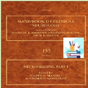
تصویربرداری عصبیپی دی اف واقعیقیمت 1 یورو2016(کیفیت اصلی)ویرایش 1تصویربرداری عصبی، قسمت اول، متنی از کتاب راهنمای عصبشناسی بالینی، نشان میدهد که چگونه تصویربرداری عصبی به سرعت در حال گسترش دامنه و کاربردهای خود در عصبشناسی بالینی است. این یک منبع ایده آل برای هر کسی است که علاقه مند به مطالعه سیستم عصبی است و هم برای مبتدیان در زمینه های مختلف مرتبط و هم برای متخصصانی که می خواهند پایگاه دانش خود را در مورد تصویربرداری عصبی به روز کنند یا به روز کنند مفید است.این جلد اول به طور خاص توصیفی از تکنیک های تصویربرداری مورد استفاده در مغز بزرگسالان را پوشش می دهد، با هدف ارائه دیدگاهی جامع از زمینه تصویربرداری عصبی برای مخاطبان مختلف. این پوشش گسترده ای از موضوع را با استفاده از بسیاری از تصاویر رنگی برای نشان دادن نکات کلیدی به ارمغان می آورد.مشارکتهای کارشناسان برجسته جهانی جمعآوری شدهاند و گستردهترین دیدگاه تصویربرداری عصبی را بهصورت فعلی ارائه میکنند. برای تعدادی از اختلالات عصبی، تصویربرداری نه تنها برای تشخیص مهم است، بلکه برای نظارت بر اثر درمان ها نیز ضروری است و کل این زمینه از درمان بیماری ها به پیشگیری از آنها در حال حرکت است. بیشتر اطلاعات موجود در این جلد نشان دهنده جدید بودن این رویکرد است و به این افق جدید در مطالعه اختلالات عصبی اشاره می کند.شرح مرتبطی از فناوریهای مورد استفاده در تصویربرداری عصبی، از جمله توموگرافی کامپیوتری (CT)، تصویربرداری تشدید مغناطیسی (MRI)، توموگرافی گسیل پوزیترون (PET) و چندین مورد دیگر را ارائه میکند.منبع ایده آل برای هر کسی که سیستم عصبی را مطالعه می کند، از مبتدی تا متخصصی که علاقه مند به پیشرفت های اخیر در تصویربرداری عصبی از مغز بزرگسالان است.در مورد کاربرد تکنیک های تصویربرداری در مطالعه بیماری های مغز و نخاع و استفاده از آن در سندرم های مختلف بحث می کند.
-
درباره نویسندهجوزف سی. ماسدو، دکترای دکترا، دارای کرسی ممتاز خانواده گراهام در علوم عصبی در مؤسسه پزشکی آکادمیک متدیست هیوستون است و مرکز ملی آلزایمر و تصویربرداری عصبی نانتز را در مؤسسههای تحقیقاتی و عصبی متدیست هوستون رهبری میکند. او استاد نورولوژی در کالج پزشکی ویل کورنل است.دکتر Masdeu به استفاده از ابزارهای تصویربرداری عصبی، از جمله نشانگرهای تصویربرداری عصبی مولکولی جدید، برای مطالعه اختلالات عصبی مغز علاقه مند است. این تحقیق با شفاف سازی مکانیسم های بیماری و ارائه نشانگرهای قابل سنجش پیشرفت بیماری، منجر به تولید داروهای جدید می شود و آزمایش درمان های جدید را در نمونه های کوچکتر و با مشخصات بهتر بیماران تسهیل می کند.دکتر ماسدو که بومی اسپانیا است و پس از مدرک دکترای خود در دانشگاه مادرید در رشته روانپزشکی گواهینامه دریافت کرد، یک شهروند ایالات متحده است که دوره های رزیدنتی را در شیکاگو به پایان رساند و در سال 1976 توسط هیئت عصب شناسی و روانپزشکی آمریکا گواهینامه نورولوژی دریافت کرد. او در سال های 1976-1977 در بیمارستان زنان و بریگهام دانشکده پزشکی هاروارد در رشته آسیب شناسی اعصاب مشغول به کار بود. در دهه 1980، در کالج پزشکی آلبرت انیشتین، در نیویورک، بخش تحقیقاتی آلزایمر را در پروژه برنامه ای در مورد پیری و زوال عقل هدایت کرد. از سال 1991 تا 2000 او رئیس بخش نورولوژی کالج پزشکی نیویورک بود. او از سال 2000 تا 2008 استاد نورولوژی و مدیر علوم اعصاب در دانشکده پزشکی دانشگاه ناوارا در پامپلونای اسپانیا و پزشک و دانشمند ارشد در بخش تصویربرداری عصبی یکپارچه شاخه اختلالات مغزی بالینی برنامه تحقیقات درون مدرسه ای بود. مؤسسه ملی بهداشت (NIMH) از 2008-2014. او نویسنده 142 مقاله بررسی شده، 61 فصل کتاب، و هفت کتاب، از جمله “محلی سازی در عصب شناسی بالینی” است که اکنون در ویرایش ششم آن منتشر شده است. او با علاقه دیرینه به تصویربرداری عصبی، رئیس گروه تحقیقاتی تصویربرداری عصبی فدراسیون جهانی نورولوژی است. او مدیر آکادمی نورولوژی آمریکا و رئیس انجمن تصویربرداری عصبی آمریکا بوده است. از سال 2007 او سردبیر مجله تصویربرداری عصبی است.دکتر آر. او دکترای خود را از دانشکده پزشکی هاروارد و دکترای خود را در شیمی بیوفیزیک از دانشگاه کالیفرنیا در سانتا کروز دریافت کرد. او کار پس از دکترا را در بیوفیزیک مولکولی در آزمایشگاه ملی مگنت فرانسیس بیتر در MIT انجام داد. آموزش بالینی او شامل دوره کارآموزی در پزشکی و رزیدنتی در رادیولوژی در بیمارستان بریگهام و زنان در بوستون و فلوشیپ نورورادیولوژی در MGH بود. او بیش از 200 مقاله علمی معتبر و معتبر، بیش از 100 فصل و نقد و 3 کتاب منتشر کرده است. او در زمینههای سکته مغزی ایسکمیک، تومورهای مغزی، خونریزی مغزی، زوال عقل ایدز و بیماری آلزایمر مقالاتی منتشر کرده است و بودجه تحقیقاتی را از NIH، بنیادها و شرکتهای پزشکی دریافت کرده است. او بیش از 150 سخنرانی دعوت شده در دانشگاه ها و نشست های علمی در ایالات متحده، اروپا، آمریکای لاتین و آسیا داشته است. او افتخارات متعددی از جمله جایزه پژوهشگر برجسته از انجمن رادیولوژی اعصاب آمریکا دریافت کرده است و سخنرانی های Taveras (هاروارد)، نیوتن (UCSF) و وود (UNC) را ارائه کرده است. او درخواست های کمک مالی را به عنوان عضوی از بخش های مطالعاتی NIH و چندین سازمان علمی بررسی کرده است. دکتر گونزالس عضو هیئت تحریریه مجله آمریکایی نور رادیولوژی و جراحی مغز و اعصاب بوده است و داور موقت بسیاری از مجلات از جمله مجله پزشکی نیوانگلند، لانست، رادیولوژی و سکته مغزی است. او با همسرش میشل، شیمیدان اتمسفر، و دو دخترشان سوزان و آنا در کمبریج زندگی می کند.
-
فهرستتصویربرداری توموگرافی کامپیوتری و آنژیوگرافی – اصولتصویربرداری MR: ساختارشکنی نمودارهای زمان بندی و ابهام زدایی از فضای kروش های حجمی و ردیابی فیبر MRI برای ماده خاکستری و سفیدتصویربرداری رزونانس مغناطیسی عملکردیطیف سنجی رزونانس مغناطیسی بالینی سیستم عصبی مرکزیپرفیوژن مغز: توموگرافی کامپیوتری و تکنیکهای تشدید مغناطیسیآنژیوگرافی رزونانس مغناطیسی: اصول و کاربردهای فیزیکیآنژیوگرافی تشخیصی عروق مغزی نخاعینورسونولوژی و تصویربرداری غیر تهاجمی از شریان های کاروتیدمیلوگرافی: تکنیک مدرن و نشانه هاتوموگرافی گسیل پوزیترونتوموگرافی انتشار پوزیترون: تصویربرداری لیگاندتوموگرافی تابشی تک فوتونیتومورهای مغزی داخل محوریتومورهای مغزی خارج محوریتصویربرداری از سکته مغزی ایسکمیک حادسایر بیماری های انسداد عروق مغزیبیماری هموراژیک عروق مغزیعفونتاسکلروز چندگانهسایر اختلالات التهابی غیر عفونیتصویربرداری از ضربه به سراختلالات مخچه: یافته های بالینی/رادیولوژیک و ابزارهای تصویربرداری مدرنتصویربرداری از اختلالات ژنتیکی و دژنراتیو که در درجه اول باعث پارکینسونیسم می شونداختلالات ژنتیکی و دژنراتیو در درجه اول باعث سایر اختلالات حرکتی می شونداختلالات ژنتیکی و دژنراتیو در درجه اول باعث زوال عقل می شوندسندرم های عصبی پوستیجریان مایع مغزی نخاعی در بزرگسالاناختلالات متابولیک ارثی یا اکتسابیتصویربرداری از ضایعات قاعده جمجمهتصویربرداری از اختلالات اربیتال
شامل تصاویر پر جنب و جوش و رنگارنگ برای نشان دادن نکات کلیدی است
 How to Buy
Neuroimaging TRUE PDF price 1€
you should pay 1€ Euro via below payment Link
https://yekpay.io/en/baharmedbook.com
Pay attention to Send Money To abbas kouhiesfahani baharmedbook_manager_Bank account
After Payment confirmation ,contact us by shopping guide on WhatsApp or info@baharmedbook.com
and You will Receive Direct Download Link
———————————————————
Product Description
———————————————————
Download e-book
———————————————————
Neuroimaging
TRUE PDF
Price 1€
2016
(Original quality)
1th Edition
Neuroimaging, Part One, a text from The Handbook of Clinical Neurology illustrates how neuroimaging is rapidly expanding its reach and applications in clinical neurology. It is an ideal resource for anyone interested in the study of the nervous system, and is useful to both beginners in various related fields and to specialists who want to update or refresh their knowledge base on neuroimaging.
This first volume specifically covers a description of imaging techniques used in the adult brain, aiming to bring a comprehensive view of the field of neuroimaging to a varying audience. It brings broad coverage of the topic using many color images to illustrate key points.
Contributions from leading global experts are collated, providing the broadest view of neuroimaging as it currently stands. For a number of neurological disorders, imaging is not only critical for diagnosis, but also for monitoring the effect of therapies, and the entire field is moving from curing diseases to preventing them. Most of the information contained in this volume reflects the newness of this approach, pointing to this new horizon in the study of neurological disorders.
- Provides a relevant description of the technologies used in neuroimaging, including computed tomography (CT), magnetic resonance imaging (MRI), positron emission tomography (PET), and several others
- Ideal resource for anyone studying the nervous system, from beginners to specialists interested in recent advances in neuroimaging of the adult brain
- Discusses the application of imaging techniques to the study of brain and spinal cord disease and its use in various syndromes
- Contains vibrant, colorful images to illustrate key points
About the Author
Joseph C. Masdeu, MD, PhD holds the Graham Family Distinguished Endowed Chair in Neurological Sciences at the Houston Methodist Institute of Academic Medicine and leads the Nantz National Alzheimer Center and Neuroimaging at the Houston Methodist Neurological and Research Institutes. He is Professor of Neurology at Weill Cornell Medical College.
Dr. Masdeu is interested in the application of neuroimaging tools, including new molecular neuroimaging markers, to the study of neurodegenerative brain disorders. By clarifying disease mechanisms and providing quantifiable markers of disease progression, this research will lead to the production of new medications and greatly facilitate the testing of new therapies in smaller and better characterized patient samples.
A native of Spain, where he became certified in Psychiatry after his MD degree at the University of Madrid, Dr. Masdeu is a US citizen who completed residency training in Chicago and was certified in Neurology by the American Board of Neurology and Psychiatry in 1976. He was a fellow in Neuropathology at the Brigham and Women’s Hospital of Harvard Medical School in 1976-77. In the 1980s, at the Albert Einstein College of Medicine, in New York, he directed the Alzheimer research component of a Program Project on Aging and Dementia. From 1991-2000 he was the Chairman of Neurology at the New York Medical College. He was Professor of Neurology and Director of Neurosciences at the University of Navarra Medical School, in Pamplona, Spain, from 2000-2008 and a Senior Staff Physician and Scientist at the Section of Integrative Neuroimaging of the Clinical Brain Disorders Branch of the Intramural Research Program of the National Institutes of Health (NIMH) from 2008-2014. He is the author of 142 peer-reviewed papers, 61 book chapters, and of seven books, including “Localization in Clinical Neurology,” now in its 6th edition. With a longstanding interest in neuroimaging, he is chairman of the Neuroimaging Research Group of the World Federation of Neurology. He has been a director of the American Academy of Neurology and president of the American Society of Neuroimaging. Since 2007 he is the Editor-in-Chief of the Journal of Neuroimaging.
Dr. R. Gilberto González is Chief of Neuroradiology at the Massachusetts General Hospital and Professor of Radiology at the Harvard Medical School. He received his MD from Harvard Medical School and a PhD in biophysical chemistry from the University of California at Santa Cruz. He did post doctoral work in molecular biophysics at the Francis Bitter National Magnet Lab at MIT. His clinical training included an internship in medicine and a residency in radiology at the Brigham and Women’s Hospital in Boston, and a neuroradiology fellowship at the MGH. He has published over 200 original, peer-reviewed scientific papers, over 100 chapters and reviews, and 3 books. He has published in the areas of ischemic stroke, brain tumors, cerebral hemorrhage, AIDS dementia and Alzheimer’s disease, and has received research funding from the NIH, foundations and medical enterprises. He has given over 150 invited lectures at universities and at scientific meetings in the US, Europe, Latin America and Asia. He has received several honors including the Outstanding Researcher Award from the American Society of Neuroradiology, and has given the Taveras (Harvard), Newton (UCSF) and Wood (UNC) Lectures. He has reviewed grant applications as a member of NIH study sections and for several scientific organizations. Dr. González has been on the editorial board of the American Journal of Neuroradiology and Neurosurgery and is an ad hoc reviewer for many journals including the New England Journal of Medicine, The Lancet, Radiology and Stroke. He lives in Cambridge with his wife Michele, an atmospheric chemist, and their 2 daughters Susan and Ana.
Contents
Computed tomography imaging and angiography – principles
MR imaging: deconstructing timing diagrams and demystifying k-space
Volumetric and fiber-tracing MRI methods for gray and white matter
Functional magnetic resonance imaging
Clinical magnetic resonance spectroscopy of the central nervous system
Brain perfusion: computed tomography and magnetic resonance techniques
Magnetic resonance angiography: physical principles and applications
Diagnostic angiography of the cerebrospinal vasculature
Neurosonology and noninvasive imaging of the carotid arteries
Myelography: modem technique and indications
Positron Emission Tomography
Positron emission tomography: ligand imaging
Single-photon emission tomography
Intra-axial brain tumors
Extra-axial brain tumors
Imaging acute ischemic stroke
Other cerebrovascular occlusive disease
Hemorrhagic cerebrovascular disease
Infection
Multiple sclerosis
Other noninfectious inflammatory disorders
Imaging of head trauma
Cerebellar disorders: clinical/radiologic findings and modem imaging tools
Imaging of genetic and degenerative disorders primarily causing Parkinsonism
Genetic and degenerative disorders primarily causing other movement disorders
Genetic and degenerative disorders primarily causing dementia
Neurocutaneous syndromes
Cerebrospinal fluid flow in adults
Inherited or acquired metabolic disorders
Imaging of skull base lesions
Imaging of orbital disorders