
توضیح محصول
———————————————————
دانلود کتاب الکترونیکی
———————————————————
 How to Buy
Medical Imaging TRUE PDF price 1€
you should pay 1€ Euro via below payment Link
https://yekpay.io/en/baharmedbook.com
Pay attention to Send Money To abbas kouhiesfahani baharmedbook_manager_Bank account
After Payment confirmation ,contact us by shopping guide on WhatsApp or info@baharmedbook.com
and You will Receive Direct Download Link
———————————————————
Product Description
———————————————————
Download e-book
———————————————————
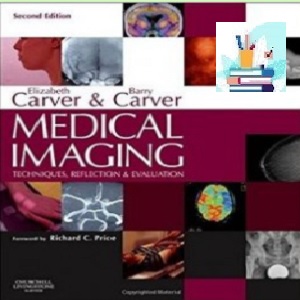
Medical Imaging
TRUE PDF
Price 1€
2013
(Original quality)
2th Edition
Medical Imaging has been revised and updated to reflect the current role and responsibilities of the radiographer, a role that continues to extend as the 21st century progresses. This comprehensive book covers the full range of medical imaging methods/techniques which all students and professionals must understand, and discusses them related to imaging principles, radiation dose, patient condition, body area and pathologies.
There is comprehensive, up-to-date, referencing for all chapters, with full image evaluation criteria and a systematic approach to fault recognition for all radiographic projections. Highly respected editors, Elizabeth and Barry Carver, have brought together an impressive team of contributing authors, comprising academic, radiographer and radiologist clinical experts.
NEW TO THIS EDITION
Full colour, including approximately 200 new colour photographs
All techniques have been updated to reflect the use of digital image receptors
All chapters have been updated to reflect current practice, eg CT colonoscopy is now included as part of GI imaging; the nuclear medicine chapter now introduces hybrid imaging; the genitourinary chapter now reflects the use of ultrasound and CT
‘The authors have been comprehensive, thorough and innovative. This well-presented book should be adopted by Schools of Diagnostic Imaging in Europe and elsewhere and be a constant companion to the reflective radiographic practitioner.’ From the foreword to the first edition by Patrick Brennan.
Full colour, including approximately 200 new colour photographs.All techniques have been updated to reflect the use of digital image receptors. All chapters have been updated to reflect current practice, eg CT colonoscopy is now included as part of GI imaging; the nuclear medicine chapter now introduces hybrid imaging; the genitourinary chapter now reflects the use of ultrasound and CT.
Elizabeth Carver, BSc(Hons), FAETC, DCRR, Undergraduate Course Director, School of Radiography, University of Wales, Bangor, UK; and Barry Carver, PgDipCT, PGCE, DCRR, Senior Postgraduate Tutor, School of Radiography, University of Wales, Bangor, Wales –This text refers to an out of print or unavailable edition of this title.
۱ Digital imaging
۲ Film/screen imaging
۳ Exposure factorsr manipulation and dose
۴ Introduction to skeletal,, chest and abdominal radiography
۵ Fingers, hand and wrist
۶ Forearm, elbow and humerus
۷ The shoulder girdle
۸ Footr toesr ankler tibia and fibula
۹ Knee and femur
۱۰ Pelvis and hips
۱۱ Cervical spine
۱۲ Thoracic spine
۱۳ Lumbar spine
۱۴ Sacrum and coccyx
۱۵ Thoracic skeleton
۱۶ Principles of radiography of the head
۱۷ Cranial vault
۱۸ Facial bones
۱۹ Paranasal sinuses
۲۰ Specialised projections of the skull
۲۱ Dental radiography
۲۲ Orthopantomography and cephalometry
۲۳ Chest and thoracic contents
۲۴ Abdomen
۲۵ Accident and emergency
۲۶ Breast imaging
۲۷ Paediatric imaging in general radiography
۲۸ Contrast media
۲۹ Gastrointestinal tract
۳۰ Accessory organs of the gastrointestinal tract
۳۱ Investigations of the genitourinary tract
۳۲ Cardiovascular system
۳۳ Vascular imaging of the head and neck
۳۴ Interventional and therapeutic procedures
۳۵ Computed tomography
۳۶ Magnetic resonance imaging
۳۷ Nuclear medicine imaging
۳۸ Ultrasound