
توضیح محصول
———————————————————
دانلود کتاب الکترونیکی
———————————————————
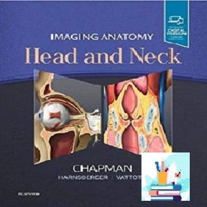
تصویربرداری آناتومی سر و گردن
پی دی اف واقعی
قیمت 1 یورو
2019
(کیفیت اصلی)
ویرایش 1
ساختارهای بسیار تخصصی، میکروآناتومی اجزای جداگانه و تراکم ساختاری کلی، سر و گردن را به یکی از چالش برانگیزترین مناطق در رادیولوژی تبدیل کرده است. تصویربرداری آناتومی: سر و گردن به رادیولوژیستها، ساکنان و همکاران یک مرجع آناتومی واقعاً جامع و فوقالعاده نشان داده شده است که برای بهبود مهارتهای تفسیری در این منطقه پیچیده طراحی شده است. انبوهی از تصاویر مقطعی با کیفیت بالا، تصاویر پزشکی مربوطه، و متن مختصر و توصیفی فرصتی منحصر به فرد برای تسلط بر اصول آناتومی طبیعی و تشخیص دقیق و کارآمد شرایط پاتولوژیک ارائه می دهد.
حاوی بیش از 1400 تصویر با وضوح بالا و مقطعی سر و گردن همراه با بیش از 200 تصویر پزشکی پر جنب و جوش، طراحی شده برای ارائه پاسخ های سریع رادیولوژیست پرمشغله به سوالات آناتومی تصویربرداری
منعکس کننده درک جدید آناتومی به دلیل تحقیقات در حال انجام آناتومیک و همچنین تکنیکهای تصویربرداری جدید و پیشرفته
دارای ویژگیهای ۳ سکانس تصویربرداری تسلا MR و توالیهای آناتومی طبیعی CT چند آشکارساز در سراسر کتاب، ارائه نماهای دقیق از ساختارهای آناتومیک که تکمیل کننده تصاویر پزشکی بسیار دقیق و دقیق است.
شامل مجموعههای تصویربرداری از برشهای متوالی در هر صفحه استاندارد تصویربرداری (کرونال، ساژیتال و محوری)
تغییرات آناتومیک و فرآیندهای پاتولوژیک را به تصویر میکشد تا به شما کمک کند ظاهر و ارتباط مورفولوژی تغییر یافته را سریع تشخیص دهید.
شامل تصاویر CT و MR از شرایط پاتولوژیک، در صورت لزوم، زیرا به طور مستقیم درک فعلی آناتومی طبیعی را افزایش میدهند.
حاوی بخش جداگانهای درباره آناتومی سونوگرافی طبیعی سر و گردن است
مرور
“به طور کلی، این مرجع عالی برای آناتومی پیچیده سر و گردن است.”
-Nikhil Maduripan، MD (مرکز پزشکی Baystate) برای خدمات بررسی Doody
فهرست
فصل 1. نمای کلی پایه جمجمه
فصل 2. قاعده جمجمه قدامی
فصل 3. پایه جمجمه مرکزی
فصل 4. قاعده جمجمه خلفی
فصل 5. آناتومی استخوان تمپورال
فصل 6. آناتومی اصلاح مورب استخوان تمپورال
فصل 7. آناتومی کانال شنوایی خارجی
فصل 8. آناتومی ماستوئید گوش میانی
فصل 9. آناتومی گوش داخلی
فصل 10. آناتومی راس سنگی
فصل 11. آناتومی CPA-IAC
فصل 12. مفصل گیجگاهی فکی
فصل 13. بررسی اجمالی Cranial Neves
فصل 14. CNI (نوعی بویایی)
فصل 15. CNII (نور نوری)
فصل 16. CNIII (نور چشمی)
فصل 17. CNIV (Trochlear Neve)
فصل 18. CNV (نف سه قلو)
فصل 19. CNVI (Abducens Neve)
فصل 20. CNVH (نور صورت)
فصل 21. CNVIII (Neve Vestibulocochlear)
فصل 22. CNIX (نور گلوسوفارنکس)
فصل 23. CNX (Vagus Neve)
فصل 24. CNXI (لوازم جانبی)
فصل 25. CNXII (Hypoglossal Neve)
فصل 26. نمای اجمالی مدار
فصل 27. مدار استخوانی و روزنه
فصل 28. کمپلکس اپتیک اپتیکال/غلاف
فصل 29. کره زمین
فصل 30. سینوس غار
فصل 31. Sinonasal Gveview
فصل 32. واحد Gstiomeatal
فصل 33. حفره پتریگوپالاتین
فصل 34. فرورفتگی پیشانی و سلول های هوای مرتبط
فصل 35. مروری بر گردن Suprahyoid و Infrahyoid
فصل 36. فضای پارافارنکس
فصل 37. فضای مخاطی حلقی
فصل 38. فضای جونده
فصل 39. فضای پاروتید
فصل 40. فضای کاروتید
فصل 41. فضای رتروفارنکس
فصل 42. فضای اطراف مهره
فصل 43. فضای گردن خلفی
فصل 44. فضای احشایی
فصل 45. حنجره
فصل 46. هیپوفارنکس
فصل 47. آناتومی تیروئید و پاراتیروئید
فصل 48. نای و مری سرویکس
فصل 49. غدد لنفاوی گردنی
فصل 50. عضلات صورت و سیستم عضلانی آپونورتیک سطحی
فصل 51. حفره دهان Gveview
فصل 52. فضای مخاطی دهان
فصل 53. فضای زیر زبانی
فصل 54. فضای زیر فکی
فصل 55. فضای باکال
فصل 56. زبان
فصل 57. تریگون رترومولار
فصل 58. مندیبل و فک بالا
فصل 59. اتصال کرانیوسوییکال
فصل 60. ستون فقرات گردنی
فصل 61. شبکه بازویی

How to Buy
Imaging Anatomy Head and Neck TRUE PDF price 1€
you should pay 1€ Euro via below payment Link
https://yekpay.io/en/baharmedbook.com
Pay attention to Send Money To abbas kouhiesfahani baharmedbook_manager_Bank account
After Payment confirmation ,contact us by shopping guide on WhatsApp or info@baharmedbook.com
and You will Receive Direct Download Link
———————————————————
Product Description
———————————————————
Download e-book
———————————————————
Imaging Anatomy Head and Neck
TRUE PDF
Price 1€
2019
(Original quality)
1th Edition
Highly specialized structures, microanatomy of individual components, and overall structural density make the head and neck one of the most challenging areas in radiology. Imaging Anatomy: Head and Neck provides radiologists, residents, and fellows with a truly comprehensive, superbly illustrated anatomy reference that is designed to improve interpretive skills in this complex area. A wealth of high-quality, cross-sectional images, corresponding medical illustrations, and concise, descriptive text offer a unique opportunity to master the fundamentals of normal anatomy and accurately and efficiently recognize pathologic conditions.
- Contains more than 1400 high-resolution, cross-sectional head and neck images combined with over 200 vibrant medical illustrations, designed to provide the busy radiologist rapid answers to imaging anatomy questions
- Reflects new understandings of anatomy due to ongoing anatomic research as well as new, advanced imaging techniques
- Features ۳ Tesla MR imaging sequences and state-of-the-art multidetector CT normal anatomy sequences throughout the book, providing detailed views of anatomic structures that complement highly accurate and detailed medical illustrations
- Includes imaging series of successive slices in each standard plane of imaging (coronal, sagittal, and axial)
- Depicts anatomic variations and pathological processes to help you quickly recognize the appearance and relevance of altered morphology
- Includes CT and MR images of pathologic conditions, when appropriate, as they directly enhance current understanding of normal anatomy
- Contains a separate section on normal ultrasound anatomy of the head and neck
Review
“Overall, this is a great reference for complex head and neck anatomy.”
-Nikhil Madhuripan, MD (Baystate Medical Center) for Doody’s Review Service
Contents
Chapter 1. Skull Base Overview
Chapter 2. Anterior Skull Base
Chapter 3. Central Skull Base
Chapter 4. Posterior Skull Base
Chapter 5. Temporal Bone Anatomy
Chapter 6. Temporal Bone Oblique Reformation Anatomy
Chapter 7. External Auditory Canal Anatomy
Chapter 8. Middle Ear-Mastoid Anatomy
Chapter 9. Inner Ear Anatomy
Chapter 10. Petrous Apex Anatomy
Chapter 11. CPA-IAC Anatomy
Chapter 12. Temporomandibular Joint
Chapter 13. Cranial Neves Overview
Chapter 14. CNI (Olfactory Neve]
Chapter 15. CNII (Optic Neve]
Chapter 16. CNIII (Oculomotor Neve]
Chapter 17. CNIV (Trochlear Neve]
Chapter 18. CNV (Trigeminal Neve]
Chapter 19. CNVI (Abducens Neve]
Chapter 20. CNVH (Facial Neve]
Chapter 21. CNVIII (Vestibulocochlear Neve]
Chapter 22. CNIX (Glossopharyngeal Neve]
Chapter 23. CNX (Vagus Neve]
Chapter 24. CNXI (Accessory Neve]
Chapter 25. CNXII (Hypoglossal Neve]
Chapter 26. Orbit Oveview
Chapter 27. Bony Orbit and Foramina
Chapter 28. Optic Neve/Sheath Complex
Chapter 29. Globe
Chapter 30. Cavernous Sinus
Chapter 31. Sinonasal Gveview
Chapter 32. Gstiomeatal Unit
Chapter 33. Pterygopalatine Fossa
Chapter 34. Frontal Recess and Related Air Cells
Chapter 35. Suprahyoid and Infrahyoid Neck Oveview
Chapter 36. Parapharyngeal Space
Chapter 37. Pharyngeal Mucosal Space
Chapter 38. Masticator Space
Chapter 39. Parotid Space
Chapter 40. Carotid Space
Chapter 41. Retropharyngeal Space
Chapter 42. Perivertebral Space
Chapter 43. Posterior Cevical Space
Chapter 44. Visceral Space
Chapter 45. Larynx
Chapter 46. Hypopharynx
Chapter 47. Thyroid and Parathyroid Anatomy
Chapter 48. Cevical Trachea and Esophagus
Chapter 49. Cevical Lymph Nodes
Chapter 50. Facial Muscles and Superficial Musculoaponeurotic System
Chapter 51. Oral Cavity Gveview
Chapter 52. Oral Mucosal Space
Chapter 53. Sublingual Space
Chapter 54. Submandibular Space
Chapter 55. Buccal Space
Chapter 56. Tongue
Chapter 57. Retromolar Trigone
Chapter 58. Mandible and Maxilla
Chapter 59. Craniocevical Junction
Chapter 60. Cevical Spine
Chapter 61. Brachial Plexus